Research Paper:
Effects of TND1128 (a 5-deazaflavin derivative),with self-redox ability,as a mitochondria activator on the mouse brain slice andits comparison with b-NMN
Authors:
Yoshihisa Kudo (Department of Anesthesiology, Hachioji Medical Center, Tokyo Medical University),
Nanae Takahashi (MD, Department of General Medicine, Hachioji Medical Center, Tokyo Medical University)
Comparison of the Effects of NMN and Deazaflavin
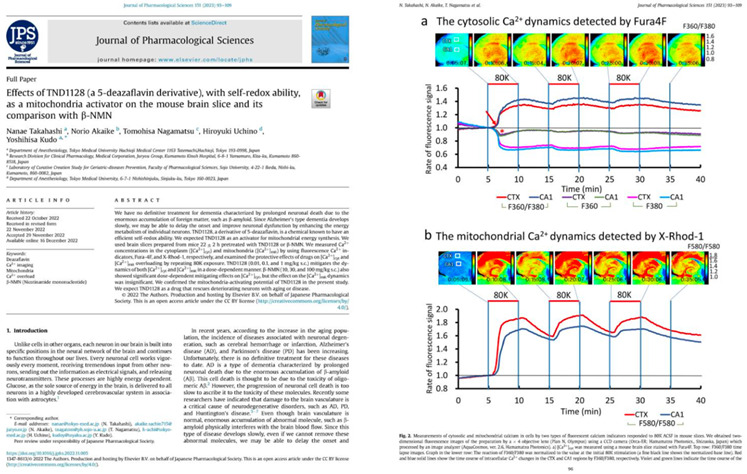
In this study, we compared the effects of β-NMN and 5-deazaflavin. Using mouse brain slice samples, we evaluated and compared their inhibitory effects on the fluctuations of Ca2+ concentration within the cytoplasm and mitochondria when exposed to depolarization stimuli.
The experiment for determining the effects was conducted 24 hours after administration, based on data indicating a significant increase in the Sir-1 gene at 24 hours post-administration.
The results showed that both β-NMN and 5-deazaflavin demonstrated the ability to control the concentration of Ca within the cytoplasm and mitochondria. However, it was found that the effect of 5-deazaflavin was more than 100 times stronger.
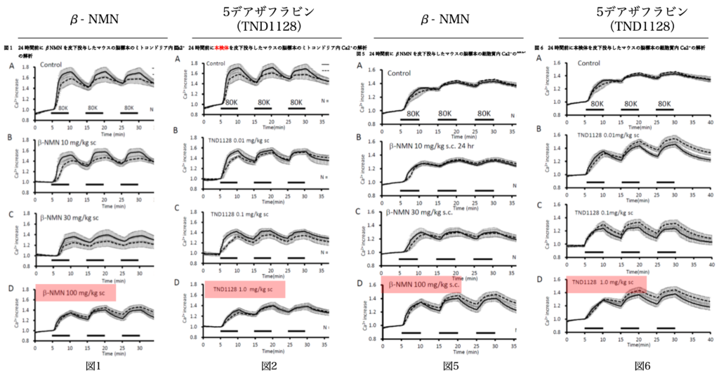
As shown in Figures 1, 2, and Figures 5, 6, examining the actions of β-NMN and deazaflavin within the cytoplasm and mitochondria reveals that they can maintain highly stable physiological responses. If this can be replicated in the human brain, it could be expected to have a powerful brain protective effect. Deazaflavin, being a highly hydrophobic and extremely stable compound, is considered for future applications.
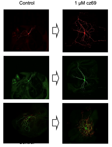
Deazaflavin activates ATP production and promotes the development of cultured hippocampal cells in the mouse brain.
Immunostaining Image of Hippocampal Neuron Axons
On the fourth day of culture, axons were immunostained with tau antibody. The left shows the Control (control group), and the right shows neuronal cells treated with Deazaflavin (1 μM) added once only on the first day of culture.
Immunostaining Image of Hippocampal Neuron Dendrites
On the fourth day of culture, dendrites were immunostained with MAP2 antibody. The left shows the Control (control group), and the right shows neuronal cells with Deazaflavin (1 μM) added once only on the first day of culture.
Immunostaining Image of Synapses in Octopus Specimens
The left shows the Control (control group), and the right shows neuronal cells where Deazaflavin (1 μM) was added once only on the first day of culture and on the fourteenth day, excitatory synapses were immunostained with VGLUT1 antibody.
Research Paper


Contact
Contact
For questions regarding products and services,
Please feel free to contact us here.

Warning: Undefined variable $official_url in /home/r2091360/public_html/kiranjapan.com/wp-content/themes/karin/fixed-cta.php on line 20
Warning: Undefined variable $amazon_url in /home/r2091360/public_html/kiranjapan.com/wp-content/themes/karin/fixed-cta.php on line 20
Warning: Undefined variable $rakuten_url in /home/r2091360/public_html/kiranjapan.com/wp-content/themes/karin/fixed-cta.php on line 20